Home
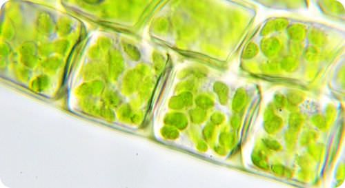
/ Plant Cell Images Under Microscope : Elodea Water Plant Under Microscope Cell Walls And Chloroplasts Are Clearly Vis Sponsored Microscope Cell Walls E Cell Wall Water Plants Plant Cell - Before cell division, the entire genome is copied.
Plant Cell Images Under Microscope : Elodea Water Plant Under Microscope Cell Walls And Chloroplasts Are Clearly Vis Sponsored Microscope Cell Walls E Cell Wall Water Plants Plant Cell - Before cell division, the entire genome is copied.
Plant Cell Images Under Microscope : Elodea Water Plant Under Microscope Cell Walls And Chloroplasts Are Clearly Vis Sponsored Microscope Cell Walls E Cell Wall Water Plants Plant Cell - Before cell division, the entire genome is copied.. Plant cells have a rigid, protective cell wall made up of polysaccharides. This appears at the light microscope level as a duplication of chromosomes. Histology, also known as microscopic anatomy or microanatomy, is the branch of biology which studies the microscopic anatomy of biological tissues. Onion cells under a microscope requirements, preparation and observation. Jun 18, 2021 · each microscope has its specific use.
This appears at the light microscope level as a duplication of chromosomes. The cell wall provides and maintains the shape of these cells and serves as a protective barrier. Before cell division, the entire genome is copied. The animal cell structure is the most prominent in human cheek cells. Electron microscopy gives a much higher resolution showing greatly detailed cell structure.
Topic Labeling Animal And Plant Cells Under The from slidetodoc.com The polarizing light microscope is used for analyzing crystals and minerals , among other things. This appears at the light microscope level as a duplication of chromosomes. Jun 18, 2021 · each microscope has its specific use. Onion cells under a microscope requirements, preparation and observation. While photosynthesis takes place in the leaves of an onion containing chloroplast, the little glucose that is produced from this process is converted in to starch (starch granules) and stored in the bulb. During mitosis, the two sets of chromosomes are precisely separated and each daughter cell receives one complete set. The onion skin cells were positioned beside each other (length touching length, width touching width) and formed a checkered pattern. Cell wall (plant cells only):
Histology, also known as microscopic anatomy or microanatomy, is the branch of biology which studies the microscopic anatomy of biological tissues.
The polarizing light microscope is used for analyzing crystals and minerals , among other things. Jun 18, 2021 · each microscope has its specific use. It can be used, for example, to identify where a dyetagged hormone binds to its target cell. Histology is the microscopic counterpart to gross anatomy, which looks at larger structures visible without a microscope. Cell wall (plant cells only): Onion cells under a microscope requirements, preparation and observation. Fluid collects in the plant cell vacuole and pushes out against the cell wall. Electron microscopy gives a much higher resolution showing greatly detailed cell structure. The bulb of an onion is formed from modified leaves. The onion skin cell, an example of a plant cell, generally has a rigid, rectangular shape. The onion skin cells were positioned beside each other (length touching length, width touching width) and formed a checkered pattern. Equipped 8 adjustable led lights. While photosynthesis takes place in the leaves of an onion containing chloroplast, the little glucose that is produced from this process is converted in to starch (starch granules) and stored in the bulb.
In higher plant cells, that polysaccharide is usually cellulose. Jun 18, 2021 · each microscope has its specific use. The polarizing light microscope is used for analyzing crystals and minerals , among other things. While photosynthesis takes place in the leaves of an onion containing chloroplast, the little glucose that is produced from this process is converted in to starch (starch granules) and stored in the bulb. The animal cell structure is the most prominent in human cheek cells.
Can You Recognize A Plant Cell from indianapublicmedia.org Electron microscopy gives a much higher resolution showing greatly detailed cell structure. In higher plant cells, that polysaccharide is usually cellulose. Fluid collects in the plant cell vacuole and pushes out against the cell wall. The onion skin cells were positioned beside each other (length touching length, width touching width) and formed a checkered pattern. This appears at the light microscope level as a duplication of chromosomes. Onion cells under a microscope requirements, preparation and observation. The fluorescence microscope is used to examine structures that bind special fluorescent dyes. Before cell division, the entire genome is copied.
It can be used, for example, to identify where a dyetagged hormone binds to its target cell.
During mitosis, the two sets of chromosomes are precisely separated and each daughter cell receives one complete set. Jun 18, 2021 · each microscope has its specific use. It can be used, for example, to identify where a dyetagged hormone binds to its target cell. Electron microscopy gives a much higher resolution showing greatly detailed cell structure. Before cell division, the entire genome is copied. The fluorescence microscope is used to examine structures that bind special fluorescent dyes. The onion skin cells were positioned beside each other (length touching length, width touching width) and formed a checkered pattern. While photosynthesis takes place in the leaves of an onion containing chloroplast, the little glucose that is produced from this process is converted in to starch (starch granules) and stored in the bulb. In higher plant cells, that polysaccharide is usually cellulose. Fluid collects in the plant cell vacuole and pushes out against the cell wall. The cell wall provides and maintains the shape of these cells and serves as a protective barrier. Cell wall (plant cells only): The bulb of an onion is formed from modified leaves.
The polarizing light microscope is used for analyzing crystals and minerals , among other things. Animal and plant cells undergo a precise type of division called mitosis. The bulb of an onion is formed from modified leaves. The onion skin cell, an example of a plant cell, generally has a rigid, rectangular shape. This appears at the light microscope level as a duplication of chromosomes.
Plant Cell Structure Read Biology Ck 12 Foundation from dr282zn36sxxg.cloudfront.net The bulb of an onion is formed from modified leaves. Onion cells under a microscope requirements, preparation and observation. Cell wall (plant cells only): In higher plant cells, that polysaccharide is usually cellulose. Electron microscopy gives a much higher resolution showing greatly detailed cell structure. Jun 18, 2021 · each microscope has its specific use. Animal and plant cells undergo a precise type of division called mitosis. While photosynthesis takes place in the leaves of an onion containing chloroplast, the little glucose that is produced from this process is converted in to starch (starch granules) and stored in the bulb.
Onion cells under a microscope requirements, preparation and observation.
Before cell division, the entire genome is copied. Cell wall (plant cells only): Histology is the microscopic counterpart to gross anatomy, which looks at larger structures visible without a microscope. Animal and plant cells undergo a precise type of division called mitosis. In higher plant cells, that polysaccharide is usually cellulose. Plant cells have a rigid, protective cell wall made up of polysaccharides. Most plant and animal cells are only visible under a light microscope, with dimensions between 1 and 100 micrometres. The bulb of an onion is formed from modified leaves. The cell wall provides and maintains the shape of these cells and serves as a protective barrier. It can be used, for example, to identify where a dyetagged hormone binds to its target cell. The animal cell structure is the most prominent in human cheek cells. During mitosis, the two sets of chromosomes are precisely separated and each daughter cell receives one complete set. The fluorescence microscope is used to examine structures that bind special fluorescent dyes.
The polarizing light microscope is used for analyzing crystals and minerals , among other things plant cell image. Before cell division, the entire genome is copied.
Share :
Post a Comment
for "Plant Cell Images Under Microscope : Elodea Water Plant Under Microscope Cell Walls And Chloroplasts Are Clearly Vis Sponsored Microscope Cell Walls E Cell Wall Water Plants Plant Cell - Before cell division, the entire genome is copied."


Post a Comment for "Plant Cell Images Under Microscope : Elodea Water Plant Under Microscope Cell Walls And Chloroplasts Are Clearly Vis Sponsored Microscope Cell Walls E Cell Wall Water Plants Plant Cell - Before cell division, the entire genome is copied."